![<?echo $_SERVER['SERVER_NAME'];?>](/template/twentyseventeen/skin/images/header.jpg)
The Guangdong Provincial People's Hospital informed the media that the dean of the Zhuang Jian team had applied mixed reality technology to nearly 10 cases of complicated congenital heart disease surgery, and “flowers and flowers†​​in the heart-like shrubs to find narrow “small targetsâ€. This is the world's first clinical breakthrough, and it is even more proud that all three 3D application-assisted diagnostic technologies and devices are sourced from China.
Break through one
3D intraoperative navigation "shifting flowers"
The 2 month old baby Xiaoan (a pseudonym), when he was born, showed signs of gradual lung failure, poor breathing, small face purple, newborns should be "windward long", and the birth weight reached Xiaoan for 2 months but still Only 3 kg weight. He was sent to the Department of Cardiology of the People's Hospital of Guangdong Province, confirming that he suffered from a serious and complicated congenital heart disease, pulmonary atresia.
On March 27th, Xiao An became the first child in the world to receive “heart surgery†with the help of mixed reality technology.
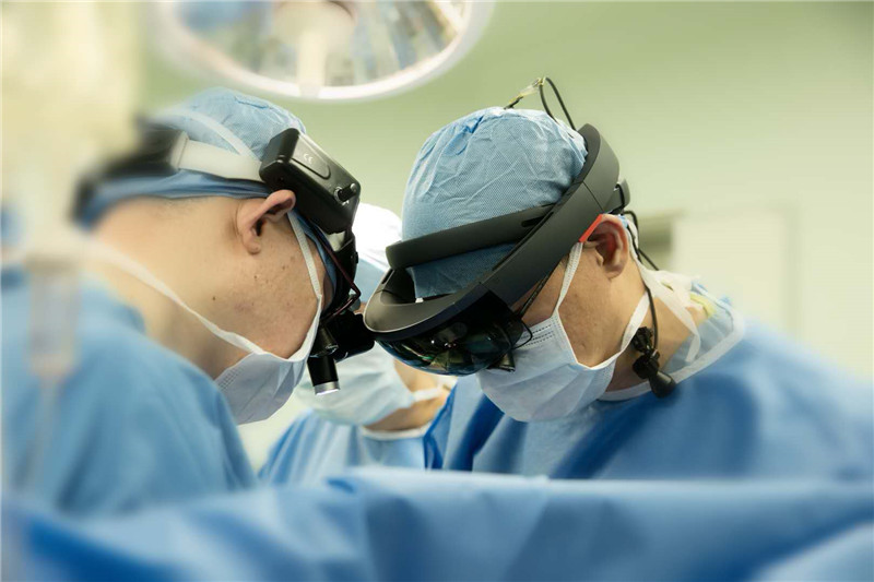
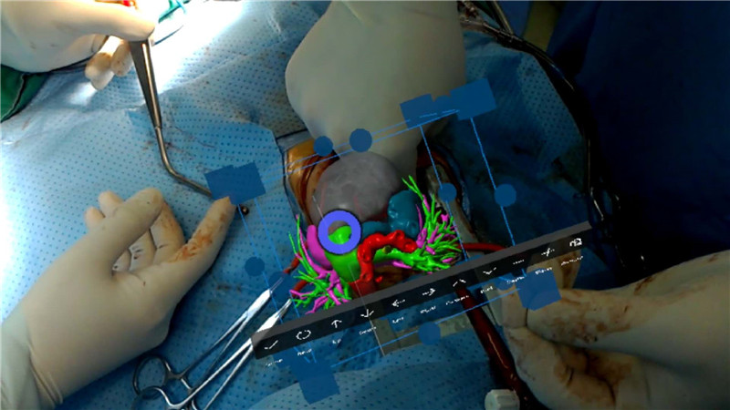
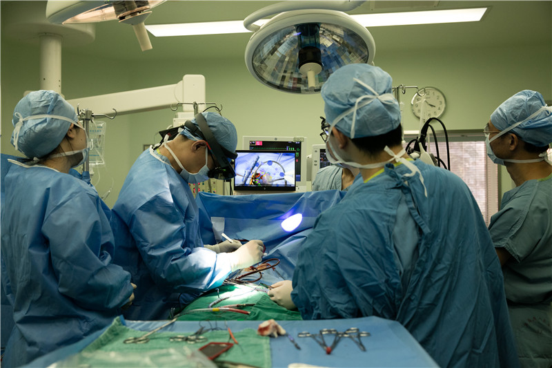
With the help of black technology MR technology, the doctors reconstructed the virtual three-dimensional Xiaoan heart image according to CT before surgery, and projected it onto the operating table. After opening the chest, the virtual heart was placed in the small chest and coincided with the actual heart. The image guides, and the strips find the bush-like collateral vessels, and then “shift the flowers†​​to where they are. The operation took only over 4 hours to complete, which was more than 6 hours shorter than usual.
Immediately after Xiao An, the 4-year-old male baby Lele (a pseudonym) was also saved. Lele is also pulmonary atresia, as well as large ventricular septal defect, patent ductus arteriosus, collateral branch of the main lung, long-term complexion, white and purple, short and fast breathing, multiple pneumonia, thin if only two years old. Lele's parents said that they have turned to a number of hospitals and said that their children's heart and lung function is too bad. They are still in depletion. They may soon be dying. It is recommended to give up. The provincial medical team is the last hope of the Lele family. Also with the help of mixed reality technology, Lele has repaired the "trunk" and "twig" of the pulmonary artery, repaired the ventricular septum, closed the catheter, and later he will grow up very close to normal children.
Decryption:
Wen Shusheng, deputy director of the provincial medical pediatric ward and deputy director of the Provincial Children's Heart Center, pointed out that pulmonary atresia like Xiaoan and Lele is a very serious congenital heart disease. The human lung care is issued by the right ventricle, just like the trunk, gradually splitting into the side branches, entering the lungs, responsible for sending the "used" hypoxic venous blood to the lungs, adding oxygen and removing carbon dioxide through breathing, becoming bright red. The arterial blood runs through the body. It is obvious how important it is to the pulmonary artery and its gradual branching!
However, the children with pulmonary atresia are equal to the absence of trunks. Those side branches can only be confused like bushes. It is long to catch a weak blood vessel. The blood supply is fragile and the efficiency is extremely low, which also causes the blood vessels in the heart to be trapped. The condition is not treated, the heart and lungs are gradually depleted, and the children often die quickly.
In the past, pulmonary atresia was relieved, and it was considered a super-surgical operation. It was necessary to open an incision in the left and right chests. A group of chaotic "shrub" collateral vessels were found, separated, and then an incision was made in the middle of the chest to splicing them one by one. On the unlocked pulmonary artery, a surgery is performed for at least 10 hours. "Going to power at 8 or 9 in the morning, to achieve 8:00 in the evening," Wen Shusheng said. "The most time-consuming and difficult thing is to find a side branch of a blood vessel. It is not a matter of finding it. It is to find the origin, direction, and free environment of each root. Etc. Hybrid MR technology is the most critical aid to solve this problem."
Break through two
Pre-operative "walking in" 3D heart hit "target"
Xiao Bing (a pseudonym), a 5-year-old girl, is also a child with complicated congenital heart disease. She had undergone heart surgery 2 years ago. Recently, she developed myocardial bundle hypertrophy and narrowing the intracardiac blood flow channel, which led to her small activities. Severe shortness of breath, growth and development are also significantly slow, and may even lead to sudden death. The team of the Provincial Out-of-Medical Comprehensive Disease Area realized that the child should be operated for the first time after diagnosis.
The doctor team firstly printed the virtual three-dimensional small ice heart reconstructed according to the CT data, which confirmed the left ventricular outflow obstruction, which was caused by the hypertrophic muscle bundle; the anastomosis of the pulmonary artery and the right ventricular outflow channel was also narrow.
On March 29, during the operation, the team projected Xiaobing's virtual 3D heart image onto the operating table. After opening the chest, the virtual heart was placed in the chest of Xiaobing, which coincided with the actual heart. The surgeon, Professor Jian Zhengzheng, also put on the surgery. The head-mounted display (VR glasses), "walking into" the inside of the virtual heart of Xiao Bing, along the blood vessels, find the deformed stenosis, "touching" the small meat ring that grows out of the vascular obstruction. With the help of three kinds of 3D technology, the team visually and intuitively found out the anatomical structure of the hypertrophic muscle bundle and the stenosis, accurately found the lesion, accurately removed the hypertrophic muscle bundle, and widened the preoperatively planned patch. In the narrow place, Xiao Bing was saved.
Decryption:
Yan Jianzheng, director of the Provincial Out-of-Medical Comprehensive Disease Area, pointed out that there are many children undergoing secondary surgery like Xiao Bing. The biggest problem is the chest that has been operated. The original layers of the tissue have been glued together. The heart of the child is already in the heart. Small, the lesions are difficult to find, there are many surrounding structures, and how many materials are used. There are many “minefields†and the risk of surgery is extremely high.
In the past, for such children, whether it is to confirm cardiovascular abnormalities, tissue identification, or specific details of surgery, they can only wait for the intraoperative exploration after opening the chest, and a little bit, the surgeon must always worry about "seeing Not clear enough."
"This kind of surgery, I used to be in the heart catheter room, I can't stay away from it." Huang Meiping, director of the provincial medical catheter room, said that the surgeon could not find out, and had to stop and nervously call her for support. CT, MRI, and heart color ultrasound Data interaction confirmed that the operation teamed up indoors and outdoors.
Now with pre-operative 3D printing, AR "walking into the heart", MR virtual heart and the actual heart "fit", all-round navigation surgery, Huang Meiping finally put down the burden of "off-site navigation."
Break through three
3D combination all realized domestically
3D printing, AR, MR, in people's eyes are "black technology", but in medical clinical applications, it is not as simple as a game to feel the virtual world.
Zhuang Jian, chairman of the Chinese Medical Association's Thoracic Heart Branch and dean of the Guangdong Provincial People's Hospital, pointed out that virtual technology is used in medical clinics. In January this year, it was reported that in December last year, international peers applied AR technology in a simple congenital treatment. The first 3D printing, AR "into the heart", MR virtual heart and the actual heart "fit" three combination of virtual technology attack, and has applied close to 10 complex congenital treatment, this is the world's first leading clinical breakthrough.
What makes Zhuang Jian even more proud is that such a global initiative, whether it is technology or equipment, is completely independent intellectual property rights in China.
He said that cardiovascular therapy, first-class treatment leadership, new technology is very valuable, because behind the strong support of patients benefit, so the provincial doctors decided to become the leader early, driving "black technology" from the experience into medical diagnosis and treatment.
The earliest virtual technology landing to the clinic is 3D printing technology . So far, a laboratory jointly established by the provincial doctor and Zhuhai has printed more than 70 virtual hearts, and realized pre-operative and intraoperative navigation after modeling. "The blood vessels are covered by tissues. In the past, half of the surgery was often found. The anatomical structure in memory could not be matched. The blood vessels could not be found, so I had to ask for help outside the court." Zhuang Jian said that there is now a virtual heart navigation. No need to worry about half-way withdrawal, extracorporeal circulation, greatly improve the safety of surgery.
VR virtual reality technology is a collaboration with Xi'an University of Electronic Science and Technology, allowing doctors to directly enter the virtual heart "heart", look for internal complex deformities, and no longer need to open the chest as before. "Cut a bit here, open everything there" In-situ exploration, "complex mind, most of them are intracardiac malformations", Zhuang Jian said, this can be too important, less damage to the patient's heart.
MR mixed reality technology is a cooperative application between a provincial doctor and a company in Heilongjiang. On the basis of augmented reality, the patient's heart image can be placed above the surgery field, and even the virtual heart and the physical heart coincide, even if it is fine. The blood vessels of the lateral branch do not need to separate the tissues everywhere, and the positioning is very precise.
Cast iron sink is made of cast iron. The exterior is applied with grade A porcelain enamel.
Cast Iron Sink,Cast Iron Bathroom Sink,Cast Iron Double Sink,Cast Iron Porcelain Sink
Anping Sunshine Sanitary Ware Co., Ltd. , https://www.sunshinebathtubs.com